Executive Summary & Key Takeaways
- Tilapia Lake Virus (TiLV) is an enveloped, negative-sense, 10-segment single-stranded RNA virus of the genus Tilapinevirus, capable of causing up to 90% cumulative mortality within 10 days of infection.
- Never confuse TiLV with bacterial Streptococcosis or Columnaris; bilateral ocular opacity (cataracts), endophthalmia, and abdominal dropsy unresponsive to antibiotics are hallmark clinical indicators.
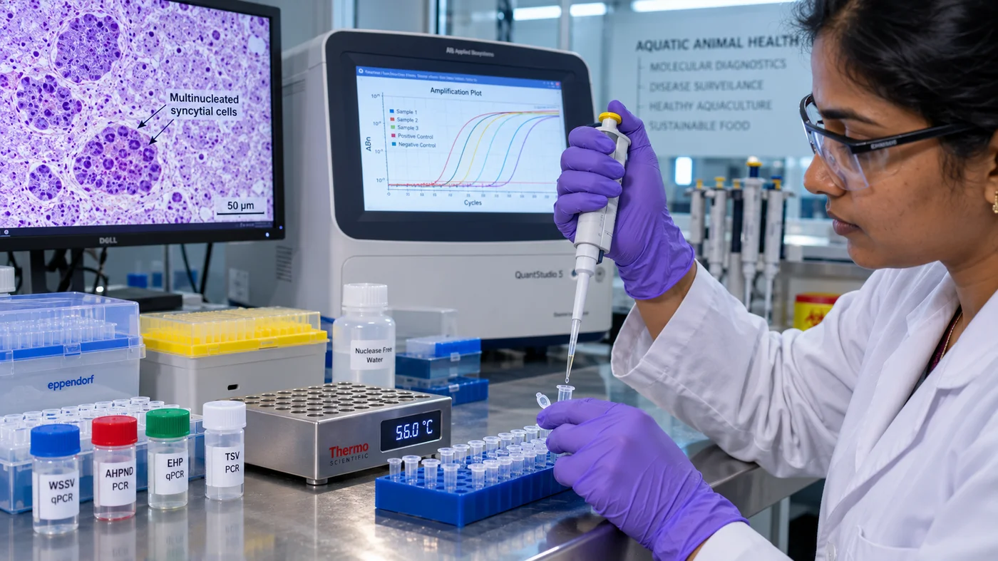
- Pathognomonic histopathology centers on multinucleated syncytial giant cell formation in hepatic parenchyma and cytoplasmic vacuolation in the brain and ocular neuroretina.
- Molecular semi-nested RT-PCR or real-time qPCR targeting genomic Segment 3 is the only definitive OIE/WOAH-approved diagnostic method for detecting subclinical TiLV carriers.
- TiLV transmits both horizontally through waterborne cohabitation and cannibalism, and vertically from asymptomatic broodstock to eggs and yolk-sac fry.
- SPF (Specific Pathogen Free) broodstock certification and 30-day quarantine isolation of new seed stocks represent the only viable commercial biosecurity shield against viral introduction.
- Upon laboratory confirmation of an outbreak, execute immediate containment: cease all water discharge, chlorinate infected tanks/ponds with active chlorine @ 30 ppm, and fallow beds under solarization for 21 days.
Clinical Field Case Study: Halting a 12-Pond Commercial Farm Outbreak
A commercial tilapia nursery and grow-out operation stocking 180,000 GIFT fingerlings across 12 semi-intensive earthen ponds experienced sudden, catastrophic lethargy and daily mortalities exceeding 2,500 fish in two nursery ponds. Affected juveniles (15g to 25g) exhibited classic clinical signs: dense bilateral corneal cataracts, swollen protruding eyes (exophthalmia), and severe dermal scale sloughing with hemorrhagic skin ulcers. Local feed suppliers suspected bacterial Streptococcosis and recommended oxytetracycline in-feed treatment, which completely failed to curb mortality. The farm engaged the AquaSangham Molecular Diagnostic Protocol: liver and brain tissue samples were harvested in RNA-preservation buffer and subjected to semi-nested RT-PCR targeting TiLV Segment 3. Within 18 hours, gels confirmed a strong 250 bp amplicon diagnostic of Tilapia Lake Virus, while histopathology revealed extensive hepatic syncytial giant cell formation. Acting decisively, the farm implemented emergency biocontainment: infected nursery ponds were isolated with zero water exchange, chemically treated with sodium hypochlorite (30 ppm active chlorine) to neutralize viral particles, and bird netting was erected to stop predatory vector transfer. Adjacent ponds were prophylactically dosed with dietary beta-glucans and sodium butyrate. The rapid molecular confirmation prevented cross-contamination across the remaining 10 grow-out ponds, saving over 140 metric tons of mature fish valued at ₹28.5 Lakhs.
1. Virology & Genome Architecture: The 10-Segment Tilapinevirus Threat
Over the past decade, the rapid global intensification of tilapia farming—spanning Nile tilapia (Oreochromis niloticus), Mozambique tilapia (O. mossambicus), blue tilapia (O. aureus), and various commercial red hybrid strains—has transformed tilapia into the second most farmed finfish group on earth, often heralded as the 'aquatic chicken'. However, this hyper-expansion suffered an unprecedented biological shock with the emergence of Tilapia Lake Virus (TiLV), an emerging viral pathogen responsible for mass mortalities ranging from 20% to over 90% in aquaculture hatcheries, intensive cage farms, biofloc tanks, and earthen grow-out ponds worldwide.
First formally identified in commercial farms and wild fisheries around Lake Kinneret (Sea of Galilee) in Israel and commercial production clusters in Ecuador, TiLV has since been clinically documented across Latin America, Africa, Southeast Asia, and throughout the major aquaculture belts of India—including Andhra Pradesh, Tamil Nadu, Kerala, and West Bengal. Officially classified by the International Committee on Taxonomy of Viruses (ICTV) as Tilapinevirus tilapiae within the family Amnoonviridae (order Articulavirales), TiLV is an enveloped, negative-sense, single-stranded RNA ((-)ssRNA) virus.
The genomic architecture of TiLV is strikingly unique and shares evolutionary affinities with the Orthomyxoviridae family (which encompasses influenza viruses and infectious salmon anemia virus / ISAV). The TiLV genome consists of exactly 10 discrete RNA segments totaling approximately 10.32 kilobases (kb) in length, ranging from Segment 1 (~1,641 nucleotides) down to Segment 10 (~465 nucleotides). Each segment contains a single open reading frame (ORF) encoding a specific structural or non-structural viral protein. Segment 1 encodes the putative RNA-dependent RNA polymerase (RdRp) catalytic subunit responsible for viral genome replication and transcription, while Segment 3 encodes a major viral core protein that serves as the universal target amplicon for international molecular diagnostic PCR surveillance.
The viral particle exhibits an icosahedral or spherical morphology measuring 55 to 75 nanometers in diameter, enveloped by a lipid bilayer membrane studded with glycoprotein spikes. Because it possesses a lipid envelope, TiLV is chemically vulnerable to lipid solvents, detergents, and common commercial aquatic disinfectants; however, within the protected aquatic environment of high-density commercial fish farms, it spreads with alarming velocity, demanding an unyielding biosecurity defense.
Taxonomic & Structural Classification
1. Family & Order: Family Amnoonviridae, Order Articulavirales, Genus Tilapinevirus.
2. Genome Dimensions: 10 negative-sense single-stranded RNA segments (10.32 kb total length).
3. Virion Morphology: Enveloped, spherical to icosahedral symmetry, 55 to 75 nm diameter.
4. Primary Target Tissues: Liver, brain (cerebral hemisphere and optic lobe), spleen, pronephros, gills, and neuroretina.
Because TiLV possesses a delicate lipid envelope, the virus is completely inactivated by exposure to temperatures above 56°C for 30 minutes, or by contact with 30 ppm free active chlorine, 100 ppm povidone-iodine, or 0.5% potassium peroxymonosulfate. Maintaining dry, solarized pond bottoms breaks viral persistence reliably.
2. Clinical Symptoms: Field Pathology Across Fry, Fingerlings & Adults
Tilapia Lake Virus affects all production stages of tilapia, but the severity of morbidity and the velocity of mortality vary considerably across life cycles. Nursery fry (0.5g to 5g) and fingerlings (5g to 30g) represent the most vulnerable demographic, where acute outbreaks often manifest as explosive mortality curves claiming 80% to 90% of the entire tank or hapa population within 5 to 10 days of the initial clinical onset. In adult grow-out fish (300g to 700g), mortality typically stabilizes between 15% and 50%, though survivors frequently suffer severe, irreversible ocular damage and somatic stunting that completely destroys commercial slaughter value.
Clinical manifestation begins with pronounced behavioral alterations. Affected fish cease voluntary feeding (anorexia), exhibit lethargic schooling near the surface or along pond dykes, lose normal rheotaxis (inability to orient against aeration currents), and eventually display erratic, uncoordinated corkscrew spinning or surface gasping as viral replication causes extensive lesions in the central nervous system.
Gross external pathological hallmarks are distinctly characteristic, though they must never be used alone for definitive diagnosis without molecular confirmation:
1. Ocular Pathology (The 'Cloudy Eye' Syndrome): Up to 80% of clinically diseased fish exhibit bilateral or unilateral ocular opacity. This manifests as dense cataracts of the lens, corneal ulceration, and severe exophthalmia ('pop-eye') or, conversely, endophthalmia (sunken, shrunken eye sockets) resulting from intraocular pressure collapse and retinal detachment.
2. Dermal Lesions & Scale Sloughing: Fish display extensive patches of epidermal erosion, loss of dermal mucus, and widespread scale loss (sloughing). The underlying dermis develops prominent petechial to ecchymotic hemorrhages, progressing to deep necrotic open skin ulcers, particularly along the operculum, ventral abdomen, and caudal peduncle.
3. Abdominal Dropsy & Fin Rot: Severe abdominal distension (ascites) filled with straw-colored serous fluid is common. The pectoral, dorsal, and caudal fins exhibit severe fraying, congestion, and hemorrhagic erosion at their bases.
Internal necropsy reveals profound systemic multi-organ pathology. Upon opening the peritoneal cavity, farm technicians will observe massive hepatomegaly; the liver appears severely swollen, friable, and discolored (ranging from pale creamy-yellow to dark mottled brownish-red with necrotic foci). The spleen is consistently enlarged (splenomegaly) and dark blackish-purple. The gastrointestinal tract is completely devoid of ingested feed, filled only with clear or bile-stained mucoid fluid, and the gall bladder is markedly distended with dark green bile.
If you observe a sudden spike in daily mortality accompanied by bilateral cloudy cataracts and fish swimming in uncoordinated circles, immediately quarantine the pond before administering any treatments. Never apply formalin, copper sulfate, or chemical parasitic baths to TiLV-infected tanks; harsh chemical treatments inflict severe respiratory stress on virally damaged gills, multiplying daily mortality tenfold.
3. Differential Diagnosis: TiLV vs Streptococcus vs Aeromonas vs Columnaris
In commercial pond and cage culture, fish health managers routinely misdiagnose TiLV outbreaks as acute bacterial infections—most commonly bacterial Streptococcosis (caused by Streptococcus iniae or Streptococcus agalactiae) or Motile Aeromonas Septicemia (MAS, caused by Aeromonas hydrophila). This misidentification leads to thousands of dollars wasted on futile oral or waterborne antibiotic treatments, during which the viral epidemic spreads unchecked across neighboring ponds and farms.
To establish prompt, accurate clinical triaging, farm managers must compare gross pathognomonic markers using the AquaSangham Differential Finfish Diagnostic Matrix:
| Clinical Parameter | Tilapia Lake Virus (TiLV) | Streptococcosis (S. iniae/agalactiae) | Motile Aeromonas Septicemia (MAS) | Columnaris (Flavobacterium) |
|---|---|---|---|---|
| Primary Causative Agent | Enveloped (-)ssRNA Virus | Gram-positive Coccobacillus | Gram-negative Bacillus | Gram-negative Filamentous Rod |
| Ocular Pathology | Bilateral cloudy cataracts, endophthalmia or pop-eye | Unilateral/bilateral exophthalmia with hyphema (bloody eye) | Occasional mild exophthalmia without lens cataracts | No ocular lesions; clear lenses |
| Skin & Scales | Extensive scale sloughing, dermal erosion, open ulcers | Darkening of body skin (melanosis), ventral reddening | Hemorrhagic petechiae on skin, scale protrusion (dropsy) | Yellow-white 'saddleback' erosions on dorsal ridge |
| Liver Gross Pathology | Enlarged, pale, friable, multinucleated syncytia | Normal to pale liver with fibrous capsular adhesions | Congested, bright hemorrhagic liver with petechiae | Normal internal organs; localized external damage |
| Brain & Neurological | Corkscrew swimming, lethargy, encephalomalacia | Violent 'C-shape' spinning, curvature of the spine | Lethargy without distinct corkscrew swimming | Normal neurological behavior until moribund |
| Antibiotic Response | ZERO response; mortality continues unabated | High clinical response to Florfenicol / Doxycycline | Moderate response to Oxytetracycline / Enrofloxacin | High response to Oxytetracycline / Potassium Permanganate |
| Definitive Diagnostic | Semi-nested RT-PCR / Real-Time qPCR (Segment 3) | Bacterial culture on Blood Agar / Strep PCR | Culture on Rimler-Shotts / Nutrient Agar | Wet mount microscopy of gill/skin scrapings (haystack motility) |
The decisive clinical test in the field is antibiotic responsiveness: If a high-mortality outbreak involving cloudy eyes and lethargic spinning fails to show a dramatic 50%+ reduction in daily mortality within 72 hours of therapeutic florfenicol administration, suspect TiLV immediately and submit tissue samples for RT-PCR confirmation.
4. Histopathology: Multinucleated Syncytial Giant Cells & Inclusions
While molecular PCR assays provide definitive genetic identification of viral nucleic acids, histopathology provides the definitive microscopic proof of active viral cytopathology in target host tissues. For veterinary pathologists and diagnostic wet-labs, TiLV exhibits an unmistakable, pathognomonic hallmark: the formation of multinucleated syncytial giant cells within the liver and brain parenchyma.
Syncytial cells develop when the fusogenic envelope glycoproteins of Tilapia Lake Virus mediate the abnormal fusion of adjacent infected host cell membranes. Instead of maintaining individual cell boundaries, dozens of infected hepatocytes coalesce into massive, multinucleated giant cells (often containing 5 to 25 aggregated nuclei clustered within a shared, amorphous eosinophilic cytoplasm).
Histopathological manifestations across specific organ systems include:
1. Hepatic Pathology: The liver is the primary epicenter of viral replication. Microscopic examination of hematoxylin and eosin (H&E) stained liver sections reveals severe dissociation of hepatic cords, widespread cytoplasmic vacuolation (hydropic degeneration), focal areas of coagulative necrosis, and prominent multinucleated syncytial giant cells. In addition, round to irregular intracytoplasmic eosinophilic inclusion bodies are frequently visible within hepatocytes and syncytia.
2. Central Nervous System (CNS) Pathology: In the brain—particularly the cerebral hemispheres, optic tectum, and cerebellum—TiLV induces severe lymphocytic meningoencephalitis. Histopathologists observe extensive perivascular cuffing (infiltration of lymphocytes around cerebral micro-vessels), focal gliosis, neuronal degeneration, and edema of the neuroretina.
3. Splenic & Pronephric Pathology: The spleen exhibits massive proliferation of melanomacrophage centers (MMCs), marked by dense accumulations of dark brownish-black hemosiderin and lipofuscin pigments. Severe lymphoid depletion is evident across both the spleen and anterior kidney (pronephros), severely impairing the fish's cellular immune defense and paving the way for opportunistic secondary bacterial invasions.
When preserving fish tissues for histopathological examination, never freeze the samples. Carefully dissect 5mm tissue slices of fresh liver, brain, and spleen from a moribund (live, dying) fish and immerse them immediately in 10% Neutral Buffered Formalin (NBF) at a 10:1 fixative-to-tissue ratio. Freezing creates ice crystal artifacts that destroy delicate syncytial cellular architecture.
5. Molecular Diagnostics: Semi-Nested RT-PCR & qPCR Protocols
Because TiLV often establishes latent, subclinical carrier states in mature broodstock and fingerlings without causing overt clinical cataracts or mortality, phenotypic inspection is completely inadequate for biosecurity clearance. The World Organisation for Animal Health (WOAH, formerly OIE) and national diagnostic reference laboratories recognize molecular nucleic acid amplification—specifically Semi-Nested Reverse Transcription Polymerase Chain Reaction (RT-PCR) and Real-Time Quantitative RT-PCR (RT-qPCR)—as the gold standard for TiLV surveillance.
Diagnostic Workflow for Semi-Nested RT-PCR targeting TiLV Segment 3:
Step 1: Tissue Harvesting & RNA Extraction. Harvest 25 to 50 mg of pooled liver, spleen, and brain tissue from suspected fish. Homogenize tissues in a dedicated lysis buffer (such as TRIzol or silica-column lysis reagents) and extract total viral RNA according to standard guanidinium thiocyanate-phenol-chloroform protocols. Quantify RNA purity on a spectrophotometer (ensuring an A260/A280 ratio between 1.8 and 2.1).
Step 2: Reverse Transcription (cDNA Synthesis). Convert extracted single-stranded viral RNA into stable complementary DNA (cDNA) using a high-fidelity reverse transcriptase enzyme (such as M-MLV or SuperScript III) primed with random hexamers or sequence-specific primers at 42°C for 50 minutes, followed by enzyme inactivation at 70°C for 15 minutes.
Step 3: Primary (First-Round) PCR Amplification. The first round of PCR utilizes specific external primers designed by Eyngor et al. to amplify a 415-base-pair (bp) fragment of TiLV genomic Segment 3:
- Forward Primer (ME1): 5'-GTT GGG CAC AAG GCA TCA TA-3'
- Reverse Primer (ME2): 5'-TAT CAC GTG GCG GAC TAA GA-3'
Reaction Cycling: Initial denaturation at 95°C for 5 min; followed by 35 cycles of denaturation at 94°C for 30 sec, primer annealing at 56°C for 30 sec, extension at 72°C for 45 sec; and a final extension at 72°C for 5 min.
Step 4: Semi-Nested (Second-Round) PCR Amplification. To achieve ultra-high diagnostic sensitivity capable of detecting as few as 10 to 50 viral genome copies in asymptomatic carriers, 1.0 µL of the first-round PCR product is transferred into a second reaction mixture containing the nested primer pair:
- Nested Forward Primer (Nested F): 5'-TAT CAC GTG GCG GAC TAA GA-3' (ME2)
- Nested Reverse Primer (Nested R): 5'-CAA TGA AAT GTT GAT CCC CAC T-3' (TiLV-Nest-R)
Reaction Cycling: 30 cycles with an annealing temperature of 58°C, yielding a sharp, definitive 250-base-pair (bp) diagnostic amplicon visualized on a 1.5% agarose gel stained with ethidium bromide or GelRed under ultraviolet transillumination.
For high-throughput commercial hatcheries and border biosecurity screening, TaqMan-based Real-Time RT-qPCR assays offer same-day quantitative results with cycle threshold (Ct) cutoffs: Samples displaying Ct values < 30 indicate high viral loads with active replication; Ct values between 30 and 38 indicate low-level asymptomatic carrier states; and Ct > 38 (or no amplification) designates the sample as TiLV-Negative.
Always include verified positive control cDNA and non-template negative control (nuclease-free water) in every PCR run. When shipping tissue samples to diagnostic testing labs, submerge organs in 95% molecular-grade ethanol or RNAlater at a 5:1 volume ratio to preserve viral RNA from enzymatic degradation during transit.
6. Transmission Vectors: Vertical Hatchery Spread & Horizontal Exposure
Understanding the epidemiological pathways of Tilapia Lake Virus is essential for erecting impenetrable farmgate biosecurity perimeters. Epidemiological field studies have definitively confirmed that TiLV transmits through two distinct biological pathways:
1. Horizontal Waterborne Transmission: In grow-out ponds, biofloc tanks, and open-water cage arrays, the primary mode of transmission is direct horizontal exposure. Infected fish shed massive quantities of viable virions into the water column through cutaneous mucus sloughing, epidermal ulcer exudates, feces, and gill secretions. Susceptible cohabitating fish contract the virus via oral ingestion of contaminated water, cannibalistic scavenging on diseased moribund carcasses, or direct virion absorption across damaged gill membranes and cutaneous micro-abrasions. Experimental waterborne challenge trials demonstrate that healthy tilapia cohabitated with TiLV-shedding fish develop clinical disease and begin dying within 5 to 10 days post-exposure.
2. Vertical & Hatchery Transmission: Perhaps the most devastating discovery in tilapia epidemiology is that TiLV is vertically transmitted from asymptomatic, carrier broodstock to their reproductive gametes, fertilized eggs, and yolk-sac fry. The virus has been molecularly detected in the ovaries, testes, follicular fluid, and seminal plasma of seemingly healthy adult breeders. Consequently, a single infected commercial hatchery shipping uncertified monosex fry or fingerlings can inadvertently seed the viral pathogen across hundreds of customer farms spanning an entire continent within a single breeding season.
3. Non-Piscine Vectors & Fomites: While tilapia species are the primary clinical hosts, wild piscivorous birds (such as cormorants, herons, and kingfishers) serve as dangerous mechanical vectors, carrying infected fish carcasses or contaminated droplet water between adjacent farm reservoirs. In addition, unsterilized harvesting seines, dip nets, transport crates, sorting tables, and boots act as common inanimate fomites that transfer live virions across pond clusters.
Never purchase tilapia fry or fingerlings from commercial hatcheries that cannot furnish a valid, batch-specific Certificate of Analysis demonstrating Negative TiLV status via RT-PCR testing. Sourcing uncertified 'cheap' seed is the direct cause of over 85% of catastrophic farm outbreaks in India.
7. Farm Biosecurity: SPF Broodstock, Quarantine Staging & Disinfection
Because there are currently no globally licensed commercial vaccines or curative pharmaceutical antiviral therapies approved for Tilapia Lake Virus in food fish, prevention through rigorous, multi-barrier biosecurity is the sole mechanism of survival for commercial enterprises.
The AquaSangham Five-Pillar TiLV Biosecurity Standard:
Pillar 1: Specific Pathogen Free (SPF) Broodstock Screening. Commercial seed producers must maintain closed-recirculation broodstock holding facilities with HEPA-filtered air and dual-stage UV/ozone water sterilization. Every prospective breeding cohort must undergo non-lethal surveillance (fin clip and blood serum RT-PCR screening) twice annually. Any individual or hapa testing positive for TiLV RNA must be culled immediately.
Pillar 2: Mandatory 30-Day Seed Quarantine Staging. When sourcing fingerlings from external sources, never introduce new arrivals directly into commercial grow-out ponds or cages. Transfer incoming fish into dedicated, isolated quarantine tanks situated at least 100 meters away from production facilities. Maintain fingerlings at 28°C to 30°C for a minimum of 21 to 30 days under close clinical observation. On Day 14 and Day 28, randomly sample 30 fish for pooled RT-PCR testing. If the cohort remains symptom-free and PCR-negative, they may be cleared for grow-out stocking.
Pillar 3: Water Intake Biosecurity & Disinfection. Wild aquatic fauna carry latent viral reservoirs. Raw surface water drawn from rivers, canals, or estuarine estuaries must pass through multi-stage intake filtration (100-mesh nylon filter bags) into a sealed reservoir. Water must be aged and disinfected with sodium hypochlorite (@ 25 to 30 ppm active chlorine) or potassium permanganate (@ 4 to 6 ppm) and aerated for 48 hours to neutralize residual chlorine before distribution into culture tanks.
Pillar 4: Physical Barrier Defenses. Enclose intensive nursery tanks and tarpaulin biofloc clusters under steel-framed polyhouse canopies equipped with 50% shade netting and anti-bird wire grids. For open earthen ponds, install tight overhead monofilament bird scaring lines (spaced 3 meters apart) and perimeter crab fencing (0.5-meter high smooth plastic barrier) to block ambulatory wildlife vectors.
Pillar 5: Equipment Sterilization & Footbaths. Each pond or tank cluster must maintain dedicated nets, secchi disks, feed buckets, and sampling gear. Never share unsterilized equipment between ponds. Install vehicle wheel-wash dips and personnel footbaths containing 200 ppm iodophor or 1.0% Virkon Aquatic at every farm entrance, refreshing the chemical disinfectant twice weekly.
Install dedicated, clearly marked red dip nets and sampling buckets for each individual pond or tank. Storing equipment in a 100 ppm povidone-iodine immersion barrel between samplings completely eliminates mechanical cross-contamination.
8. Outbreak Containment: Depopulation, Chlorination & Fallow Protocols
When a farm experiences a laboratory-confirmed TiLV outbreak, half-measures and hesitation lead to financial ruin. Immediate, uncompromising emergency biocontainment must be executed within 6 hours of diagnosis to prevent the epidemic from engulfing adjacent production units and regional watersheds.
The Emergency TiLV Outbreak Containment SOP:
Phase 1: Immediate Hydraulic Lockout. Completely seal all inlet and outlet drainage gates of the affected pond. Disconnect overflow pipes and halt all water exchange immediately. Under no circumstances should infected or moribund water be discharged into public irrigation canals or river estuaries; doing so violates national biosecurity regulations and infects neighboring aquaculture farms.
Phase 2: Humane Depopulation & Bio-Disposal. Moribund and surviving fish within the infected pond must be rapidly harvested and humanely depopulated. Dead carcasses must never be sold to local wet markets, fed to carnivorous fish, or dumped along pond dykes where birds and stray animals can feed upon them. Carcasses must be incinerated on-site or buried in a deep, sealed containment trench (minimum 1.5 meters depth) lined with a 10 cm layer of unslaked quicklime (calcium oxide, CaO) and backfilled with compact clay soil.
Phase 3: Total Chemical Sterilization of Pond Water. Prior to water discharge, the entire standing water volume of the affected pond must be chemically sterilized to eradicate all live virions. Dissolve technical-grade Calcium Hypochlorite (bleaching powder, 65% available chlorine) at a rate of 30 to 35 ppm active chlorine (equivalent to 45 to 55 kg of bleaching powder per 1,000 m³ water). Maintain continuous aeration for 24 hours to ensure thorough chemical distribution throughout the water column and benthic boundary layer. Allow the water to stand undisturbed for 7 days until residual chlorine dissipates naturally to 0.0 ppm, verified by DPD chemical titration, before discharging.
Phase 4: Deep Sludge Removal, Liming & Solarization Fallow. Drain the sterilized water and mechanically excavate the top 10 to 15 cm of black benthic organic sludge from the pond bottom. Broadcast agricultural quicklime (CaO) across the entire pond bed and dykes at a heavy rate of 1,000 to 1,500 kg per hectare. The hydration of quicklime generates intense thermal heat and drives soil pH above 11.5, saponifying any residual viral envelopes. Fallow the dry pond bed under direct tropical sunlight for a minimum of 21 to 30 days until deep, gaping desiccation cracks (10 to 15 cm deep) form across the entire floor.
Phase 5: Digital Reporting & Biosecurity Logging. Log the outbreak timeline, diagnostic PCR gel images, and containment steps directly within the AquaSangham Disease Radar & Diagnostic Suite. Rapid incident mapping alerts regional aquaculture clusters, enabling neighboring farmers to enact preemptive biosecurity lockdowns and preserve the collective biosecurity of the aquaculture ecosystem.
After chemical sterilization and fallowing, conduct a preliminary sentinel bioassay before restocking the entire farm. Stock 200 certified SPF fingerlings into a small hapa net inside the refilled pond for 14 days. If the sentinel fish show 100% survival, active feeding, and test negative on follow-up RT-PCR, the pond bed is certified pathogen-free and safe for commercial repopulation.
Summary Operational Action Checklist
Frequently Asked Questions
Q: Is Tilapia Lake Virus (TiLV) dangerous to humans or can it be transmitted to consumers?
No. Tilapia Lake Virus (TiLV) is strictly an aquatic animal pathogen that infects cichlid fish species (Oreochromis niloticus, O. mossambicus, and commercial hybrids). It is completely non-zoonotic and possesses zero infectivity or health risk to humans, whether through handling, water contact, or consuming properly cooked fish. However, diseased fish with ocular cataracts, open skin ulcers, and secondary bacterial infections are unmarketable and rejected by commercial food safety authorities.
Q: Can antibiotics like Oxytetracycline or Florfenicol cure a TiLV infection?
No. Antibiotics have zero biological efficacy against viruses. TiLV is an enveloped RNA virus that replicates inside host cells; administering antibiotics to viral infections is completely ineffective, wastes significant capital, creates antimicrobial resistance (AMR), and causes unnecessary hepatic and renal stress in already weakened fish. Antibiotics should only be prescribed if a certified veterinary lab confirms a secondary opportunistic bacterial co-infection (such as Aeromonas or Streptococcus).
Q: What is the incubation period of TiLV and how quickly does mortality occur?
Under commercial farm water temperatures (26°C to 31°C), the incubation period following horizontal waterborne exposure ranges from 5 to 10 days. Once clinical symptoms appear (anorexia, ocular cataracts, skin erosion), mortality accelerates rapidly, with peak daily losses occurring between Day 7 and Day 12. Cumulative mortality can reach 80% to 90% in juvenile populations within a single two-week window if immediate containment is not enacted.
Q: Why is Segment 3 targeted for molecular PCR diagnosis of TiLV?
The TiLV genome contains 10 single-stranded RNA segments. Segment 3 encodes a major viral core structural protein that exhibits exceptionally high sequence conservation across geographically diverse isolates from Asia, Africa, and the Americas. Validated primers targeting Segment 3 (such as ME1/ME2 and nested primers) provide the highest diagnostic specificity and analytical sensitivity, reliably detecting as few as 10 to 50 viral genome copies.
Q: Can surviving fish that recover from a TiLV outbreak be used as broodstock?
Absolutely not. Surviving tilapia that recover from an acute TiLV outbreak frequently become persistent, asymptomatic lifetime carriers. These fish harbor latent viral particles in their liver, spleen, and reproductive organs (testes and ovaries), shedding virions into the water column and passing the pathogen vertically to their fertilized eggs. Using outbreak survivors as broodstock guarantees recurring disease outbreaks in future nursery crops.
AquaSangham Technical Advisory
Aquatic Animal Health & Molecular Diagnostics Desk
Contributing Senior Technical Writer & Aqua Consultant at AquaSangham.
Recommended Farming Guides
Total Ammonia Nitrogen (TAN) Mass Balance Math: Sizing Biofilters in Commercial RAS
Read Article →Farming TechThe Recirculating Aquaculture System (RAS) Master Guide: Design, Hydraulics & Mass Balance
Read Article →Farming TechIndigenous Magur (Clarias batrachus) Induced Breeding & Nursery Rearing SOPs
Read Article →Get Live Farm Advice on AquaSangham Mobile App
Download India's #1 Aquaculture Super App for real-time market prices, precision tools, and community advice.
Download Free Android App